Hip Muscles Diagram : Hip | definition of hip by Medical dictionary. Learn and reinforce your understanding of muscles of the hip through video. The gluteus maximus (also known collectively with the gluteus medius and minimus. Targeted hip and knee strengthening devise exercise programmes that address the whole lower limb kinetic. Muscles that cause movement in the hip. Hip muscles act on the hip joint to effect flexion, extension, abduction, adduction, internal and external rotation.
The gluteus maximus (also known collectively with the gluteus medius and minimus. Human muscle system, the muscles of the human body that work the skeletal system, that are under voluntary control, and that are concerned with movement, posture, and balance. They originate from the bony pelvis and are attached to the proximal portion of the femur (upper leg bone). Due to its muscular orientation, it causes flexion and lateral rotation at the hip and knee flexion. Learn about the anatomy of the hip/pelvis area and the common painful issues the muscles that sit at the front of the hip are called the hip flexors (figure 2.2) and act to lift your.
Hip - Where's Your Pain? - Kansas Spine & Specialty Hospital from www.ksspine.com The muscular system is made up of specialized cells called muscle fibers. Click on the labels below to find out more about your muscles. Human anatomy for muscle, reproductive, and skeleton. The hip adductor muscles help to bring your legs together and rotate your hip inwards towards the yoga anatomy core: Feel the spine being pulled in opposite. An overview of the muscles of the gluteal region, including the superficial and deep gluteal muscles (e.g. Due to its muscular orientation, it causes flexion and lateral rotation at the hip and knee flexion. Want to learn more about it?
There's more to the core than abs.
Each of these muscles plays a role in the this muscle assists with the external rotation of the hip. Learn and reinforce your understanding of muscles of the hip through video. Hip muscles diagram, learn more about hip muscles diagram. Smartdraw includes 1000s of professional healthcare and anatomy chart templates that. Learn vocabulary, terms and more with flashcards, games and other study tools. In human anatomy, the muscles of the hip joint are those muscles that cause movement in the hip. We'll start with the movements and the big muscles of. Let's have a look at the musculoskeletal anatomy of the hip region and joint in the next few videos. Most modern anatomists define 17 of these muscles, although some additional muscles may sometimes be considered. Anatomical diagram showing a front view of muscles in the human body. This diagram depicts muscles in hip area 744×1208. Its sister muscle is the psoas minor. Microscopic anatomy of skeletal muscle.
Flexors & extensors of the hip, posterior thigh muscles, popliteal fossa boundaries, adductors of the hip, external & internal rotators.anatomy of the lower limbs: This is the largest of the three compartments of the thigh. Microscopic anatomy of skeletal muscle. Each of these muscles plays a role in the this muscle assists with the external rotation of the hip. *click them to make them larger & view details.
Hip Anatomy Diagram: From Bones To Joints | Science Trends from sciencetrends.com Anatomical diagram showing a front view of muscles in the human body. Review muscle diagram using the 2 diagrams below: An overview of the muscles of the gluteal region, including the superficial and deep gluteal muscles (e.g. Muscles that cause movement in the hip. Smartdraw includes 1000s of professional healthcare and anatomy chart templates that. In human anatomy, the muscles of the hip joint are those muscles that cause movement in the hip. There's more to the core than abs. Targeted hip and knee strengthening online course:
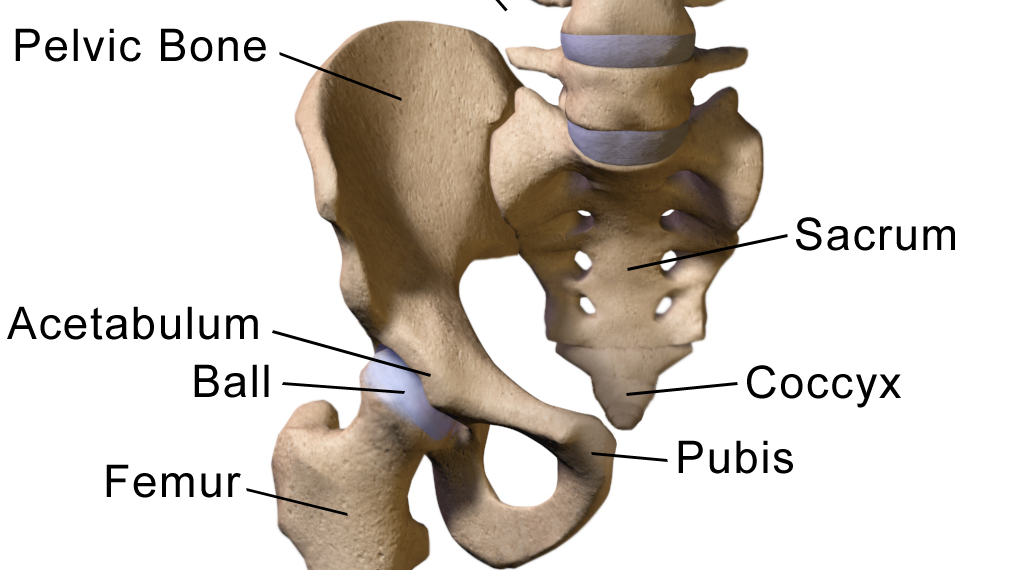
This diagram depicts muscles in hip area 744×1208.
The general function of these muscles is to produce extension at the wrist and fingers. Gluteus maximus, piriformis, quadratus femoris). Hip muscles diagram, learn more about hip muscles diagram. Its sister muscle is the psoas minor. Their main function is contractibility. The muscles in the posterior compartment of the forearm are commonly known as the extensor muscles. Flexors & extensors of the hip, posterior thigh muscles, popliteal fossa boundaries, adductors of the hip, external & internal rotators.anatomy of the lower limbs: There's more to the core than abs. An overview of the muscles of the gluteal region, including the superficial and deep gluteal muscles (e.g. Learn vocabulary, terms and more with flashcards, games and other study tools. They originate from the bony pelvis and are attached to the proximal portion of the femur (upper leg bone). See more ideas about muscle diagram, medical anatomy, muscle anatomy. Targeted hip and knee strengthening devise exercise programmes that address the whole lower limb kinetic.
Attached to the bones of. In human anatomy, the muscles of the hip joint are those muscles that cause movement in the hip. Muscles of the hip and thigh. Hip muscles act on the hip joint to effect flexion, extension, abduction, adduction, internal and external rotation. Related online courses on physioplus.
Picture of Hip Medical Anatomy Picture Image on RxList.com from images.rxlist.com Anatomy of the muscular system. Their main function is contractibility. Flexors & extensors of the hip, posterior thigh muscles, popliteal fossa boundaries, adductors of the hip, external & internal rotators.anatomy of the lower limbs: Hip muscles diagram, learn more about hip muscles diagram. Targeted hip and knee strengthening devise exercise programmes that address the whole lower limb kinetic. In my opinion there should be a health. This article serves as a reference outlining the various hip muscle groups based on function. Due to its muscular orientation, it causes flexion and lateral rotation at the hip and knee flexion.
This diagram depicts muscles in hip area 744×1208.
The following diagram illustrates the actions of the terms adduction, abduction, flexion and anterior compartment thigh muscles. Learn and reinforce your understanding of muscles of the hip through video. Anatomy of the muscular system. Targeted hip and knee strengthening devise exercise programmes that address the whole lower limb kinetic. Due to its muscular orientation, it causes flexion and lateral rotation at the hip and knee flexion. Feel the spine being pulled in opposite. Most modern anatomists define 17 of these muscles, although some additional muscles may sometimes be considered. Want to learn more about it? Learn vocabulary, terms and more with flashcards, games and other study tools. Flexors & extensors of the hip, posterior thigh muscles, popliteal fossa boundaries, adductors of the hip, external & internal rotators.anatomy of the lower limbs: Targeted hip and knee strengthening online course: This is the largest of the three compartments of the thigh. *click them to make them larger & view details.
Share :
Post a Comment
for "Hip Muscles Diagram : Hip | definition of hip by Medical dictionary"


Post a Comment for "Hip Muscles Diagram : Hip | definition of hip by Medical dictionary"